43 colon diagram with labels
How does the bowel work? Bowel information with diagrams - Macmillan ... The small bowel is part of the digestive system. It is between the stomach and the large bowel (colon). The small bowel is between 4 and 6 metres long. It folds many times to fit inside the tummy (abdomen). It breaks down food, allowing vitamins, minerals and nutrients to be absorbed into the body. The small bowel is made up of three main parts: Digestive organs: Diagram, stomach, intestines, and more The large intestine includes the cecum, transverse colon, ascending colon, descending colon, and sigmoid colon. A small, finger-like projection from the large intestine, the appendix, can become...
Label Digestive System Diagram Printout - EnchantedLearning.com Read the definitions below, then label the digestive system anatomy diagram. anus - the opening at the end of the digestive system from which feces (waste) exits the body. appendix - a small sac located on the cecum. ascending colon - the part of the large intestine that run upwards; it is located after the cecum.

Colon diagram with labels
endocrine system diagram and labels urinary system diagram unlabeled clipart digestive human unlabelled excretory clip cliparts library Digestive System With Labels Focusing On The Colon, Rectum, And Anus digestive colon rectum anus niddk focusing nih intestine Pin On Endocrine, Urinary, Reproductive, & Embryology Systems Colon (Large Intestine): Anatomy, Function, Structure The colon is comprised of four layers of tissue, similar to other regions of the digestive tract. These include: Mucosa: This is the innermost layer and is made of simple columnar epithelial tissue, making it smooth (compared to the small intestine, which contains villi, small fingerlike protrusions). Many glands secrete mucus into the interior ... Colon Histology Slide with Labeled Diagram - AnatomyLearner Colon Histology Slide with Labeled Diagram 04/06/2022 04/06/2022 by anatomylearner The colon histology slide possesses the typical four layers of a tubular organ - mucosa, submucosa, muscularis, and serosa. But, there are no permanent plica circularis and villi in the colon slide as found in the different segments of the small intestine.
Colon diagram with labels. label diagram of liver Digestive System with Labels Focusing on the Colon, Rectum, and Anus we have 14 Pictures about Digestive System with Labels Focusing on the Colon, Rectum, and Anus like Liver Anatomy Labeled Diagram Stock Photo 220894729 : Shutterstock, Liver Diagram : Living Donor Liver Transplant : Documents similar to and also PPT - Fetal Pig Dissection ... spleen diagram labeled Dissection spleen labeled colon intestine cecum pancreas diaphragm ascending ileum ileocecal artery. Histology a464. Anatomy thoracic plate spleen diagram labeled The Image Center we have 7 Pics about The Image Center like Histology A464, Cat dissection lab_labeled_images and also Cardiac muscle. A. Longitud inal section of cardiac muscle. Blog - Create a sequence diagram This works similar to release numbering, where each lifeline adds another point. Add the message number and a colon at the start of the message label. E.g. 1.3: searchByItem(itemName) Open this sequence diagram in the diagrams.net viewer. Frame labels for sequence fragments. The type of sequence fragment is written in the top left of the frame ... Colon Picture Labeling Flashcards | Quizlet Start studying Colon Picture Labeling. Learn vocabulary, terms, and more with flashcards, games, and other study tools. Search. Create. Log in Sign up. Upgrade to remove ads. ... ch.5 pathway of food diagram 14 Terms. olivia_ba. Digestive System Trace 28 Terms. lscott3. RBCs from mitral valve to tricuspid valve->traveling thru big toe 23 Terms ...
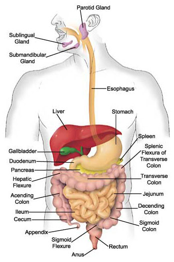
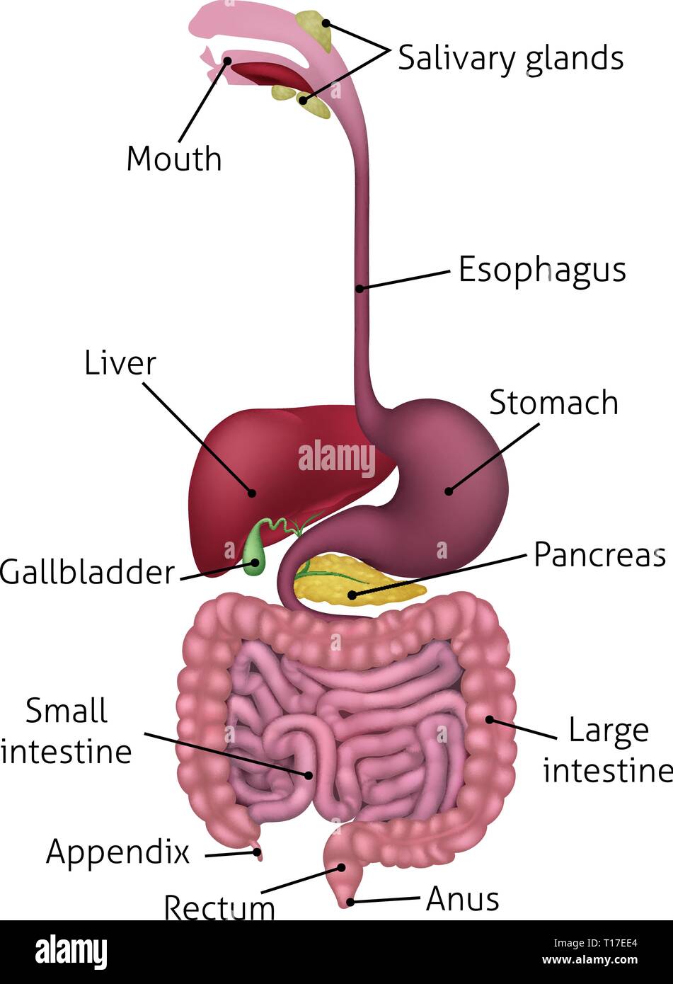
Illustration Picture of Anatomy - Colon - eMedicineHealth The colon is the largest part of the large intestine, extending from the cecum to the rectum. It is 5 feet long and its function is to reabsorb water from digested food and concentrate solid waste material, known as stool. The colon is made of several sections. The ascending colon travels up the right side of the abdomen. Understanding the Human Stomach Anatomy With Labeled Diagrams The former is located on the left, at the level of the tenth thoracic vertebra (T10). The pyloric orifice lies at the level of the first lumbar vertebra (L1). It is the opening from where the food travels towards the duodenum. Given below is a labeled diagram of the stomach to help you understand stomach anatomy. The stomach is divided into ... Colon Anatomy - Human Body Diagrams - Medical Art Library The large intestine begins at the cecum. The ileum (small intestine) ends where it connects to the cecum at the ileocecal junction. The colon is divided into four parts: the ascending, transverse, descending and sigmoid. The ascending and transverse colon meet at the right hepatic flexure (near the liver). The transverse and descending colon ... Abdomen and digestive system anatomy: diagrams labeled Full labeled anatomical diagrams - Anatomy of the abdomen and digestive system: these general diagrams show the digestive system, with the major human anatomical structures labeled (mouth, tongue, oral cavity, teeth, buccal glands, throat, pharynx, oesophagus, stomach, small intestine, large intestine, liver, gall bladder and pancreas).
Colon Anatomy (with Small Intestine Label) - NCI Visuals Online 720x602. View. Download. Title: Colon Anatomy (with Small Intestine Label) Description: Drawing shows the cecum, ascending colon, transverse colon, descending colon, sigmoid colon, rectum, and anal canal. Also shown is the small intestine. The cecum connects the small intestine to the colon. Picture of the Human Colon Anatomy & Common Colon Conditions - WebMD The ileum (last part of the small intestine) connects to the cecum (first part of the colon) in the lower right abdomen. The rest of the colon is divided into four parts: • The ascending colon... skull diagrams to label Skeletal System Diagram - Types of Skeletal System Diagrams, Examples, More. 11 Images about Skeletal System Diagram - Types of Skeletal System Diagrams, Examples, More : Skull diagram, anterior view with labels part 2 - Axial Sk… | Flickr, Human Skull Front Simplified Bones Clip Art at Clker.com - vector clip and also Human Skull Front Simplified Bones Clip Art at Clker.com - vector clip. immune system diagram without labels Skeletal System Diagram Without Labels Printable Human Skeleton Diagram we have 9 Images about Skeletal System Diagram Without Labels Printable Human Skeleton Diagram like Diagram Of The Endocrine System Without Labels - Diagram Media, Body Systems Activity Centres and also Skeletal System Diagram Without Labels Printable Human Skeleton Diagram.
Digestive tract with labels for the mouth, esophagus, stomach, small intestine, large intestine ...
female pelvis diagram labeled And Pelvis Labeled Structures Include Large Bowel Colon Diagrams Female medicinebtg.com labeled bowel medicinebtg pelvis include Cross Section At Bladder-Prostate Junction And CT prostate ct section cross pelvis bladder junction oblique axial male pricing labeled Anatomy Rendering Gallery | Kezan's Portfolio kezan.eu

Media Library | NIDDK
female pelvis diagram labeled transvaginal sagittal plane sonography anatomy pelvis diagram female normal scan uterus sonogram corpus showing figure And Pelvis Labeled Structures Include Large Bowel Colon Diagrams Female medicinebtg.com colon pelvis bowel medicinebtg urinary excretory Male Reproductive System Model Labeled - Google Search | Female
Patient Resources | Gastroenterology and Hepatology
Colon Diagram Intestine - diagrams of the large intestine colon ital is ... label the large intestine colon quiz Colon Diagram Intestine. Here are a number of highest rated Colon Diagram Intestine pictures on internet. We identified it from trustworthy source. Its submitted by doling out in the best field.

Abdomen Picture Image on MedicineNet.com
Colon: Anatomy, histology, composition, function | Kenhub The colon forms part of the large intestine and extends between the caecum and the rectum. It is about 1.5 meters in length and consists of four parts: ascending transverse descending sigmoid colon You can recognize it easily through several distinct morphological features like semilunar folds and pouches called haustra.

Gastrointestinal System
Colonoscopy Measurements (cm) from Anal Verge | SEER Training Types of Surgery: Colon; Types of Surgery: Rectum; Radiation Therapy; Commonly Used Drugs; For hands-on exercises, please go to SEER*Educate. Resources. Archived Modules. Updates. Acknowledgements. Colonoscopy Measurements (cm) from Anal Verge. Return to Anatomy of Colon and Rectum. Follow SEER. Contact Information.
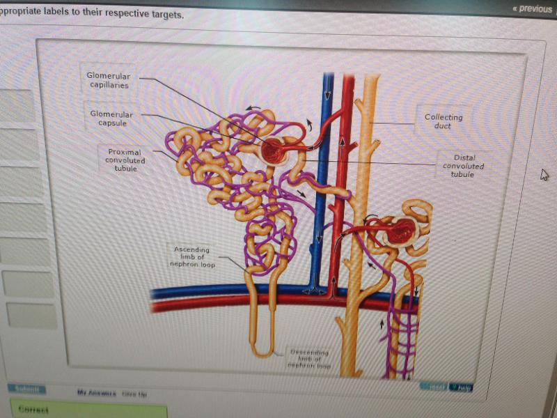
Chapter 25: The Urinary System (Mastering) Flashcards | Easy Notecards
Colon Diagram Stock Illustrations - 3,315 Colon Diagram ... - Dreamstime Download 3,315 Colon Diagram Stock Illustrations, Vectors & Clipart for FREE or amazingly low rates! New users enjoy 60% OFF. 189,239,444 stock photos online. ... Labeled Diagram. Human colon. Colon cancer. 3d rendered illustration of a transparent female anatomy with tumor in colon. Female colon. 3d rendered illustration of a transparent ...

Large Intestine Histology - Colon (labels) - histology slide - | Histology slides, Tissue ...
40 Colon diagram Vector Images, Colon diagram Illustrations - Depositphotos 40 Colon diagram Stock Vector Images, Royalty-free Colon diagram Drawings & Illustrations. VectorMine Crohns disease vector illustration. Labeled diagram with diagnosis. VectorMine Ulcerative colitis vector illustration. Labeled anatomical infographic.
Human Gastrointestinal Digestive System and Labels Stock Vector Image & Art - Alamy
Intestines (Anatomy): Picture, Function, Location, Conditions The large intestine (colon or large bowel) is about 5 feet long and about 3 inches in diameter. The colon absorbs water from wastes, creating stool. As stool enters the rectum, nerves there create ...
Post a Comment for "43 colon diagram with labels"