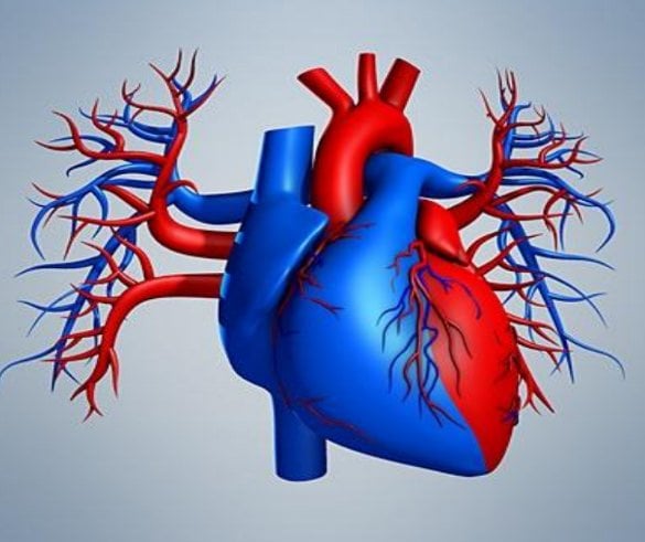
44 human heart diagram and labels
human heart drawing with colour - Good Throw Newsletter Pictures All the best Human Heart Drawing Outline 39 collected on this page. Anatomy Coloring Pages Heart. Download and print these Anatomy Heart coloring pages for free. Human anatomical heart made in graphic art as laconic logo The human heart with the arteries made in graphic style with bright purple-pink human heart drawing stock illustrations. 30 Informative Facts, Diagram & Parts Of Human Body For Kids The human blood is composed of red blood cells, white blood cells, platelets, and plasma. Up to 60% of the human body is water. A human heart beats around 100,000 times in a day. An adult heart beats around 60-80 times per minute, while a child's heart beats 100 - 120 times. Skin is the largest human organ.
Coronary arteries and cardiac veins: Anatomy and branches - Kenhub Coronary arteries and cardiac veins. The heart is a muscular, four-chambered organ that is responsible for distributing blood throughout the body. The continuous activity of the heart creates a large demand for nutrients to be delivered to cardiac tissue and for waste to be removed. However, because the organ is several layers thick, it is not feasible for the tissue to obtain nutrients from ...

Human heart diagram and labels
Heart ventricles: Anatomy, function and clinical aspects - Kenhub The heart is made up of four muscular chambers that work synergistically to propel blood throughout the body. The heart is divided into two sides by the interatrial and interventricular septa. Each half of the heart has two chambers: an atrium, and a lower chamber called the ventricle. Charts of Normal Resting and Exercising Heart Rate The heart is an organ located just behind and slightly to the left of the breastbone, and pumps blood through a network of veins and arteries known as the circulatory system. The right atrium is sent blood from the veins, and delivers it to the right ventricle. It's then pumped into the lungs where it is oxygenated. Circulatory System Diagram - New Health Advisor Coronary circuit mainly consists of cardiac veins including anterior cardiac vein, small vein, middle vein and great (large) cardiac vein. There are different types of circulatory system diagrams; some have labels while others don't. The color blue stands for deoxygenated blood while red stands for blood which is oxygenated.
Human heart diagram and labels. Heart histology: Cells and layers - Kenhub The heart is composed of left and right atria and ventricles. The atria are superior to (and less muscular than) the ventricles. The right and left atria are receptacles for blood coming from the body and lungs, respectively. On the other hand, the right and left ventricles eject blood from the heart to the lungs and body, respectively. 10+ Basic Heart Diagram | Robhosking Diagram Label main parts of heart online worksheet for high school. Source: i.pinimg.com. We hope this picture basic anatomy of heart diagram can help you study and research. Source: . Explore the heart and lungs through online science activities. Source: cliparts.co. Human heart diagram anatomy tattoo. Video: Heart and circulatory system — How they work - Mayo Clinic Video: Heart and circulatory system. Your heart is a pump. It's a muscular organ about the size of your fist and located slightly left of center in your chest. Together, your heart and blood vessels make up your cardiovascular system, which circulates blood and oxygen around your body. Your heart is divided into four chambers. Diagram of Human Heart and Blood Circulation in It Exterior of the Human Heart A heart diagram labeled will provide plenty of information about the structure of your heart, including the wall of your heart. The wall of the heart has three different layers, such as the Myocardium, the Epicardium, and the Endocardium. Here's more about these three layers. Epicardium
Circulatory system - Wikipedia The circulatory system includes the heart, blood vessels, and blood. The cardiovascular system in all vertebrates, consists of the heart and blood vessels. The circulatory system is further divided into two major circuits - a pulmonary circulation, and a systemic circulation. The pulmonary circulation is a circuit loop from the right heart taking deoxygenated blood to the lungs where it is ... Left Coronary Artery: Anatomy, Function, and Significance Anatomy. Function. Clinical Significance. The larger of the two major coronary arteries, the left coronary artery (often called the left main coronary artery) emerges from the aorta and is a primary source of blood for the ventricles and left atrium of the heart. It moves to the left, coursing between the pulmonary trunk (which divides into the ... Circulatory System: Definition, Parts, Functions, Organs The Human Circulatory system is an organ system that contains the heart and the blood vessels and moves blood throughout the body in a closed network of blood capillaries. Humans have a closed circulatory system. The human circulatory system functions to transport blood and oxygen from the lungs to the various tissues of the body. What are the 12 cranial nerves? Functions and diagram The cranial nerves are a set of twelve nerves that originate in the brain. Each has a different function responsible for sense or movement. The functions of the cranial nerves are sensory, motor ...
Labelled Heart Diagram Coronary Artery - Wiring Schematic Online In this interactive you can label parts of the human heart. Catheterization of the heart is a somewhat invasive but commonly employed procedure for visualization of the heart s coronary vessels chambers valves and or great vessels. The coronary arteries are on the heart surface left main right coronary. Anatomical Line Drawings - Medscape Surface Anatomy - lateral views - male. go to drawing without labels. Surface Anatomy - lateral views - female. go to drawing without labels. Surface Anatomy - Child - anterior view & posterior ... Blood Cell Basics - Activity - TeachEngineering Students learn about the form and function of the human heart through lecture, research and dissection. ... Using their Blood Cell Model Worksheet, have students draw a diagram of their model. Ask them to label the white and red blood cells on their diagram. Ask the students the following questions: Why are there only a few white blood cells ... Body Circulation - Lesson - TeachEngineering Students are introduced to the circulatory system, the heart, and blood flow in the human body. Through guided pre-reading, during-reading and post-reading activities, students learn about the circulatory system's parts, functions and disorders, as well as engineering medical solutions. By cultivating literacy practices as presented in this lesson, students can improve their scientific and ...

Heart Diagram Worksheet Blank Human Heart Worksheets Label Heart Diagram Worksheet Awesome in ...
Heart valves anatomy: Tricuspid-aortic-mitral-pulmonary - Kenhub The mature heart is a muscular tube that is divided into four chambers: two atria (upper chambers) and two ventricles (lower chambers). The flow of blood through the heart is partially regulated by unidirectional valves that exist between the atria and ventricles.
Heart to Heart - Lesson - TeachEngineering Identify the parts of the heart (left and right ventricles, left and right atria, interventricular septum, mitral valve, tricuspid valve, pulmonary valve, aortic valve, pericardium, valve leaflets, aorta). Describe how blood flows through the heart in a specific path. Explain how problems with the heart may cause health concerns.
Heart - Wikipedia The human heart is situated in the mediastinum, at the level of thoracic vertebrae T5-T8.A double-membraned sac called the pericardium surrounds the heart and attaches to the mediastinum. The back surface of the heart lies near the vertebral column, and the front surface sits behind the sternum and rib cartilages. The upper part of the heart is the attachment point for several large blood ...
Lymphatic System: Lymphatic Functions, Diagram at Embibe Fig: Human Lymphatic System. Lymphatic System Components A. Lymph. Lymph is a fluid connective tissue that flows inside the specialised vessels known as lymphatic vessels. It is a colourless fluid that is a part of the tissue fluid, that in turn, is a part of the blood plasma.
Cardiac muscle tissue histology | Kenhub Cardiac muscle tissue, also known as myocardium, is a structurally and functionally unique subtype of muscle tissue located in the heart, that actually has characteristics from both skeletal and muscle tissues. It is capable of strong, continuous, and rhythmic contractions that are automatically generated.
Path of Blood Through the Heart - New Health Advisor Here are the basic parts of the heart: 1. Right Atrium The heart can be divided into right and left halves, as well as into the upper and lower chambers. There are two upper chambers called atria and two lower chambers called ventricles. On each side of the heart, there are one atrium and one ventricle.
The Parts of the Heart and their Functions - Step To Health Although the cavities and valves are the best-known parts of the heart, other components are essential for this organ to function. First, there's the sinus node. The sinus node is the structure that functions as a cardiac pacemaker. That is, it generates electrical impulses that are transformed into the contractions of the heart muscles.
Heart: Anatomy | Concise Medical Knowledge - Lecturio The heart is divided into 4 chambers: 2 upper chambers for receiving blood from the great vessels, known as the right and left atria, and 2 stronger lower chambers, known as the right and left ventricles, which pump blood throughout the body. Blood flows through the heart in 1 direction, moving from the right side of the heart, through the lungs


Post a Comment for "44 human heart diagram and labels"